
An echocardiogram is one of the most commonly used diagnostic tests in cardiology. It is a safe, painless, and non-invasive imaging test that allows doctors to examine the structure and function of the heart in real time.
Using sound waves, an echocardiogram produces detailed images of the heart, helping cardiologists assess how well the heart is pumping blood and identify potential abnormalities. This test plays a crucial role in diagnosing and monitoring many different heart conditions.
If you have been advised to undergo an echocardiogram, understanding how the test works and why it is recommended can help you feel more comfortable and prepared.
What Is an Echocardiogram?
An echocardiogram, often called an “echo”, uses high-frequency sound waves (ultrasound) to create images of the heart. These sound waves bounce off the structures of the heart and are converted into images displayed on a monitor.
This imaging technique allows cardiologists to observe:
-
The size and shape of the heart
-
The movement of the heart walls
-
The function of the heart valves
-
Blood flow through the heart chambers
-
The overall pumping strength of the heart
Because echocardiography provides real-time images, it helps doctors detect problems that may not appear in other tests.
Why Is an Echocardiogram Performed?
Cardiologists may recommend an echocardiogram for several reasons. The test is commonly used to diagnose, evaluate, and monitor various heart conditions.
Some of the main reasons your doctor may order an echocardiogram include:
-
Investigating unexplained chest pain
-
Evaluating shortness of breath
-
Checking for heart valve abnormalities
-
Diagnosing heart failure
-
Detecting congenital heart defects
-
Monitoring heart function after a heart attack
-
Assessing heart murmurs
An echocardiogram can also be used to evaluate how well certain treatments or medications are working.
What Conditions Can an Echocardiogram Detect?
Echocardiography is a powerful diagnostic tool that helps identify several heart conditions, including:
Heart Valve Disease
It can detect whether heart valves are narrowed (stenosis) or leaking (regurgitation).
Heart Failure
The test measures how efficiently the heart pumps blood, often evaluated through a measurement called ejection fraction.
Cardiomyopathy
Echocardiography helps diagnose diseases that affect the heart muscle.
Congenital Heart Defects
Structural abnormalities present at birth can be detected through imaging.
Blood Clots or Fluid Around the Heart
An echocardiogram can identify fluid accumulation or clots that may affect heart function.
Types of Echocardiograms
Depending on the patient’s condition, a cardiologist may recommend different types of echocardiography.
Transthoracic Echocardiogram (TTE)
This is the most common type of echocardiogram. A handheld device called a transducer is placed on the chest to capture images of the heart.
It is completely non-invasive and usually takes about 30–45 minutes.
Stress Echocardiogram
A stress echo evaluates how the heart performs during physical activity. Images are taken before and after exercise to assess blood flow and heart function under stress.
This test is often used to detect coronary artery disease.
Transesophageal Echocardiogram (TEE)
In certain cases, a more detailed image of the heart is required. In this procedure, a probe is inserted into the esophagus to capture clearer images of the heart structures.
Because the esophagus lies close to the heart, this method provides highly detailed images.
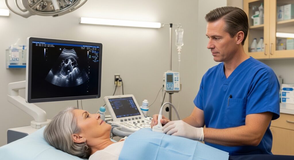
What Happens During the Test?
An echocardiogram is a simple and comfortable procedure.
During the test:
-
The patient lies on an examination table.
-
A technician applies a special gel to the chest area.
-
A small handheld device (transducer) is moved across the chest.
-
Sound waves create live images of the heart on a monitor.
The test is painless and usually takes 30 to 60 minutes to complete.
Patients can typically resume normal activities immediately after the procedure.
When Should You Consider an Echocardiogram?
Your cardiologist may recommend an echocardiogram if you experience symptoms such as:
-
Persistent chest discomfort
-
Shortness of breath
-
Irregular heartbeats or palpitations
-
Unexplained fatigue
-
Swelling in the legs or ankles
It may also be recommended if routine examinations reveal abnormal heart sounds or irregular ECG results.
Early diagnosis allows cardiologists to treat heart conditions before they become more serious.
Benefits of Echocardiography
Echocardiography offers several advantages compared to other diagnostic tests.
Some key benefits include:
-
Non-invasive and painless
-
No radiation exposure
-
Provides real-time images of the heart
-
Helps detect many heart conditions early
-
Useful for monitoring treatment progress
Because of these benefits, echocardiography is widely used as a primary diagnostic tool in cardiology.
The Importance of Early Heart Evaluation
Heart conditions can often develop gradually and may not cause noticeable symptoms in the early stages. Diagnostic tests such as echocardiography allow cardiologists to evaluate the heart’s structure and function before complications arise.
Early detection plays a crucial role in preventing serious conditions such as heart failure, severe valve disease, or cardiac arrhythmias.
Final Thoughts
An echocardiogram is a valuable and widely used diagnostic tool that helps cardiologists assess heart health with accuracy and safety. Whether used for diagnosis, monitoring, or preventive care, this imaging test provides essential insights into how the heart functions.
If you are experiencing symptoms related to heart health or have been advised by your doctor to undergo an echocardiogram, consulting a qualified cardiologist can help ensure you receive the appropriate evaluation and care.
Regular heart checkups and timely diagnostic tests can make a significant difference in maintaining long-term cardiovascular health.